A lumbar MRI specifically examines the lumbar section of your spine — the region where back problems commonly originate. The lumbosacral spine is made up of the five lumbar vertebral bones (L1 thru L5), the sacrum (the bony “shield” at the bottom of your spine), and the coccyx (tailbone).
How long does a MRI spine lumbar and sacral take?
Procedure. A lumbar spine MRI usually takes about 30-60 minutes to perform.
What can a lumbar spine MRI diagnose?
- check spinal alignment.
- detect abnormalities of vertebrae or the spinal cord.
- check for a lumbar disk herniation, which can lead to leg pain.
- evaluate any inflammation of the spinal cord or nerves.
- check for tumors on or around the spinal cord.
What does a sacral MRI show?
Used to evaluate pain in the region of the tailbone or low back pain not attributed to disorders of the lumbar spine. MRI can screen for causes of both chronic pain, as well as evaluate for fractures after episodes of trauma.Why would a doctor order an MRI of the spine?
Spine MRI detects other possible causes of back pain such as compression fracture, and bone swelling. It is also used to monitor changes in the spine after an operation, such as scarring or infection.
Do you go in feet first for a lumbar MRI?
For a cervical spine (neck), you will enter the MRI scanner head-first. For a lumbar spine, you will enter the scanner feet-first, and depending upon how tall you are, your head may be out of or near the entrance of the magnet.
Does your whole body go in for a lumbar MRI?
An MRI can be performed on any part of your body. A lumbar MRI specifically examines the lumbar section of your spine — the region where back problems commonly originate.
Will lumbar MRI show hip problems?
An MRI will often show unexpected causes of hip pain that may be originating from other nearby structures like the sacroiliac joints, pubic bones, or even the lower lumbar spine.Does an MRI show nerve damage?
An MRI may be able help identify structural lesions that may be pressing against the nerve so the problem can be corrected before permanent nerve damage occurs. Nerve damage can usually be diagnosed based on a neurological examination and can be correlated by MRI scan findings.
Is lumbar spine the same as lumbosacral spine?A lumbosacral spine x-ray is a picture of the small bones (vertebrae) in the lower part of the spine. This area includes the lumbar region and the sacrum, the area that connects the spine to the pelvis. This is the spine and the sacrum with the cervical (neck), thoracic (mid-back), and lumbar (lower back) vertebra.
Article first time published onWhat organs can be seen on a lumbar MRI?
Lumbar spine MR imaging may detect abnormalities of the kidneys, adrenal glands, liver, spleen, aorta and para-aortic regions, inferior vena cava, or the uterus and adnexal regions.
Does a lumbar MRI show the sacrum?
It relies on the use of a strong magnetic field to capture detailed pictures of the five lumbar vertebral bones, the sacrum and the coccyx (tailbone), as well as the blood vessels, tendons, nerves and ligaments that support these bones.
Does the sciatic nerve show up on an MRI?
Imaging studies are usually needed to diagnose the cause of sciatic nerve pain. An MRI of the lumbar spine will show many causes of low back pain and sciatica, including disc herniations, facet arthritis, and lumbar spinal stenosis. Digital x-rays and CT scans may also be used to diagnose the cause of sciatica.
Do you lay on your back for a spine MRI?
The MRI of the spine requires you to lie down on the scanning table.
How long does it take MRI results to come back?
The radiologist may discuss initial results of the MRI with you right after the test. Complete results are usually ready for your doctor in 1 to 2 days. An MRI can sometimes find a problem in a tissue or organ even when the size and shape of the tissue or organ looks normal.
How do I stay calm during an MRI?
- Have a family member or friend present during the MRI.
- Enjoy the warm blankets or cushions we offer. …
- You can use the lavender- and vanilla-scented eye pillows provided to help you relax and remain calm.
- Listen to music. …
- Try to control your breathing. …
- Go for a little guided mental imagery.
How long does a full spine MRI take?
Expect the whole spine MRI scan to last between 30 and 60 minutes.
What happens if you panic during an MRI?
When not properly accommodated during an MRI, claustrophobic patients may experience panic attacks, which can bring on increased heart rate, difficulty breathing, chills, sweating, and other distressing symptoms. Claustrophobia is a very common condition, affecting as much as 5% of the population.
How long does a sacral MRI take?
The test usually takes 30 to 60 minutes but can take as long as 2 hours.
How do you go through an MRI if you are claustrophobic?
- 1-Ask questions beforehand. The more educated and informed you are on the specifics of the test, the less likely you are to be surprised by something. …
- 2-Listen to music. …
- 3-Cover your eyes. …
- 4-Breathe and meditate. …
- 5-Ask for a blanket. …
- 6-Stretch beforehand. …
- 7-Take medication.
Does an MRI show arthritis?
MRI is the most effective way to diagnose problems within any joint and the image sensitivity makes it the most accurate imaging tool available in detecting arthritis and other inflammatory changes. MRI is also a key diagnostic tool when patients have lower back pain, radiating pain or hip/groin pain.
What does a pinched nerve in the lower back feel like?
Numbness or decreased sensation in the area supplied by the nerve. Sharp, aching or burning pain, which may radiate outward. Tingling, pins and needles sensations (paresthesia) Muscle weakness in the affected area.
What are the symptoms of nerve damage?
- Numbness or tingling in the hands and feet.
- Feeling like you’re wearing a tight glove or sock.
- Muscle weakness, especially in your arms or legs.
- Regularly dropping objects that you’re holding.
- Sharp pains in your hands, arms, legs, or feet.
- A buzzing sensation that feels like a mild electrical shock.
Can you see inflammation on an MRI?
MRI allows to assess the soft tissue and bone marrow involvement in case of inflammation and/or infection. MRI is capable of detecting more inflammatory lesions and erosions than US, X-ray, or CT.
Where is hip arthritis pain felt?
The pain is usually felt in the groin, but also may be felt on the side of the hip, the buttock and sometimes into the knee. Arthritis of the hip usually occurs in people as they enter their 60’s and 70’s. This varies depending on your weight, activity level and the structure of your unique hip joint.
Does lumbar spondylosis cause hip pain?
Because spinal stenosis with spondylolisthesis can cause significant hip or knee pain, the presence of degenerative arthritis at these joints must be considered. A thorough physical exam should include hip range of motion and assessment of internal and external rotation.
Is lumbar spondylosis arthritis?
Technically, spondylosis is a form of arthritis—spinal osteoarthritis (osteoarthritis is the most common type of arthritis) to be exact. We tend to think of arthritis as something you get in your hands and knees, but the spine, and all of its bones and joints, can fall victim to its grip as well.
What is Lumbo sacral pain?
Lumbosacral strain is a medical term for an injury that causes low back pain. The lumbosacral area (low back) is between the bottom of the ribcage and the top of the buttocks. A strain is tearing of muscles and tendons. These tears can be very small but still cause pain.
What does lumbo sacral mean?
Of or relating to or near the small of the back and the back part of the pelvis between the hips.
What do white spots on a spine MRI mean?
One thing that causes a lot of confusion is the presence on an MRI report of small white dots in the middle of the brain. These have a variety of names including high signal change, white matter change and small vessel disease. Sometimes they are even called ‘Unidentified Bright Objects or UBOs’.
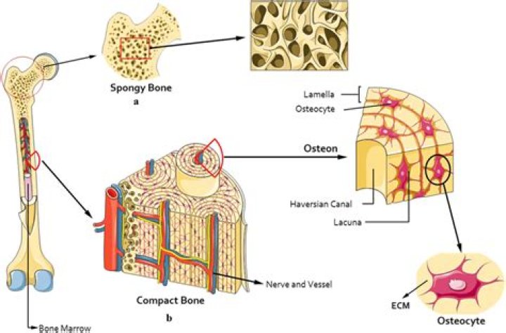
Does MRI show osteoporosis?
Other methods rather than DEXA as quantitative computed tomography and quantitative ultrasound also have a role [8]. MRI has a role in detecting osteoporosis that the appearance of bone marrow is determined by its relative amount of protein, fat, water, and cells on MRI pulse sequence.